
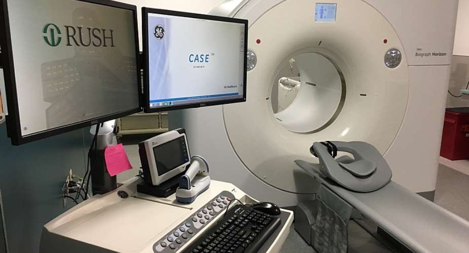
The clinical scene in nuclear medicine today is hybrid imaging. It is the modality developing most rapidly. With hybrid imaging a combination of two different modalities is meant, most commonly SPECT and computed tomography (CT) or PET and CT, resulting in a combination of molecular/biologic and structural information displayed in a fused mode (Giussani, n.d.). Hybrid systems with a combination of PET and magnetic resonance tomography (MR) are being introduced in the clinical setting, and details will be given.
The structural part, the CT, contributes with morphologic information but also with transmission data, which is used for attenuation correction of the PET. The strengths of CT are the spatial resolution and the contrast. PET and SPECT on the contrary have a relatively low spatial resolution, and the strength is the very high molar sensitivity giving the possibility to examine small changes in physiology.
PET/CT was introduced to the clinical practice in the beginning of 2000. (Giussani, n.d.). And after that a tremendous development has been taking place, Compared to SPECT, the PET scanner has a significantly higher sensitivity and a slightly better spatial resolution. Instead of using gamma emitters, positron emitters are used. A positron emitter gives away a positron from the nucleus, and since this antiparticle cannot exist, it interacts with an electron and annihilates.
The combination of PET and CT (PET/CT) is a molecular imaging modality allowing us to visualize different biochemical pathways in cancer cells. Imaging is done with 11C- or 18F-labelled tracers. The sensitivity is high, making it possible to detect picomolar and down to nanomolar concentrations of the tracer in vivo and thus to perform imaging using non-pharmacological concentrations of the substances. This counts also for SPECT and gamma camera imaging. Oncology diseases are still the major indication for PET/CT even if the use for cardiac, brain imaging and inflammation is increasing. Initially, the CT examination was mainly restricted to attenuation correction and anatomical mapping to facilitate the localization of the PET uptake. Today, the vast majority of PET/CT scanners are equipped with a state-of-the-art CT, and an increasing proportion of PET/CT studies are made with high-quality diagnostic CT. This true hybrid imaging sets demands on the physicians to have qualification in both radiology and nuclear medicine to evaluate the entire hybrid examination. Collaboration between a radiologist and a nuclear medicine specialist making the evaluation together is another working strategy
A PET-CT scan combines a CT and a PET. It gives detailed information about most cancer-reported cases globally. The CT scan takes a series of x-rays from all around your body and puts them together to create a three-dimensional (3D) image. The PET scan uses a mildly radioactive drug to show up areas of your body where cells are more active than usual (“PET - CT scan | Tests and scans | Cancer Research UK,” n.d.). PET-CT scans are for many types of cancer. They are known to give more accurate in diagnosing cancer than PET or CT scans alone.
PET-CT scans can help to diagnose cancer, evaluate how big a cancer is and whether it has spread (stage cancer), and decide whether you can have surgery to remove cancer. Moreover, it can help in decision making as to what is the best treatment for cancer. To check whether cancer has come back or better still help to plan radiotherapy treatment. A PET-CT scan can also show how well a cancer treatment is working. After you have had cancer treatment, a scan may show an area that looks like there might still be some cancer left. This might not be cancer but scar tissue leftover from cancer killed off by your treatment. A PET-CT scan can sometimes show whether this tissue is active cancer or not.
PET/CT fusion images have the potential to provide important information to guide the biopsy of a mass to active regions of the tumor and to provide better maps than CT alone. To modulate field and dose of radiation therapy, it is expected that the role of PET/CT in changing management will continue to evolve in the future and that these tools will be fundamental components of the truly “personalized medicine” we are striving to deliver. The use of these techniques can occur at the time of initial diagnosis, in assessing the early response of disease to treatment, at the conclusion of treatment, and in continuing follow-up of patients.
PET/CT is among the most widely used and well-established noninvasive tools for the diagnosis of ischemic coronary disease in the western countries. It allows assessment of the physiological relevance of coronary lesions and offers a high prognostic predictive value. Although (PET) may achieve a higher accuracy than SPECT, its use has so far often been limited to large centers. Recent advances in image processing software and the advent of hybrid scanners have paved the way for fusion of image datasets from different modalities. An ideal, noninvasive technique for the diagnosis of coronary artery disease should provide complementary information on coronary anatomy as well as on pathophysiologic lesion severity. PET/CT hybrid imaging can provide such unique information, which not only improves diagnostic assessment and risk stratification but may also guide decision making with regard to revascularization in patients with coronary artery disease.
IN SUMMARY
I suggest that, the Ghana Health service (GHS) and the Ministry of Health (MoH) should train a lot of qualified personnel to operate the use of these hybrid modalities so that people with cancer confirmed cases will be treated with accuracy and efficacy. The above summary of the PET-CT scan can help to save many lives in Ghana. Below is the demographic data of cancer reported cases in Ghana as at 2020(International Agency for Research on cancer, 2020)
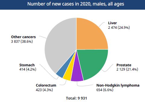
PET-CT can help us to detect the 38.6% of other cancers in the data been provide.
Emmanuel Ampofo
Level 400 radiography student at UCC
Telephone: 0246578851
E-mail: [email protected]
REFERENCING
1. Giussani, A. (n.d.). Imaging in Nuclear Medicine.
2. International Agency for Research on cancer. (2020). Ghana Cancer Fact Sheet. Global Cancer Observatory, 482, 1–2.
3. PET - CT scan | Tests and scans | Cancer Research UK. (n.d.). Retrieved August 3, 2021, from https://www.cancerresearchuk.org/about-cancer/cancer-in-general/tests/pet-ct-scan


 'We will upgrade Ho Teaching Hospital to be worthy of the name of a teaching hos...
 'If you are a man of principle and have done nothing wrong please come home' – M...
 'Ghanaians gave me one additional term; it’s exactly what the constitution says'...
 EOCO questioned Miracles Aboagye over GH¢5million, not GH¢55million – Sammi Awuk...
 'I do not take enjoyment, pride in prosecuting people if they have done no wrong...
 Father arrested for allegedly forcing her 16-year-old daughter to sleep with men...
 Female ward assistant in court over alleged GH¢1.7million military recruitment s...
 "He stood for peace, good neighborliness" — Bawku Naba mourns demise of Yaa Naa ...
 Mahama targets completion of Ho Sports Stadium before Ghana's 70th anniversary

