
When we think of Anemia, 2 structures come to mind: Hemoglobin and Red Blood Cells (Erythrocytes)
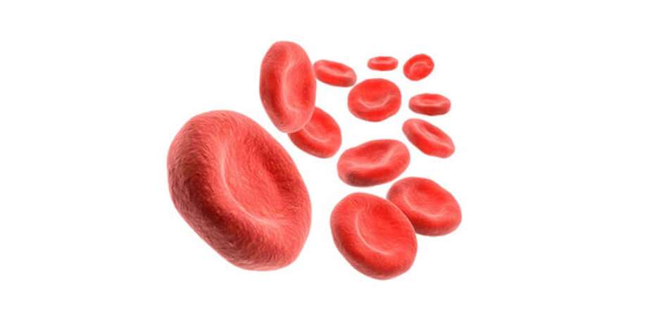
Red Blood Cells, produced continuously, in the Bone Marrow, contain Hemoglobin, the bright Red oxygen-carrier protein which gives blood its color.
Red Blood Cells (RBC) deliver Oxygen to all the body cells and tissues.
The Term Anemia is used to describe the condition, in which there is a decrease in number of circulating RBCs and/or a decrease in the hemoglobin content of the RBCs.
Anemia is unnatural, it is a sign of an underlying medical problem.
Anemia is diagnosed when the hemoglobin is <13.5 g/dl in men (<12.0g/dl in women)
CAUSES
Anemia results from one of three situations:
- . Blood Loss
- . RBC Underproduction
- . RBC Destruction
Reticulocytes, are immature RBCs freshly released from the bone marrow.
It usually takes 1 day for reticulocytes to transform into matured RBCs.
In very severe anemia, these immature RBCs could be recruited much earlier from the bone marrow, and it may take 2-3 days for them to reach maturity.
The Reticulocyte count is elevated when there is increased RBC destruction, or Blood loss.
Reticulocyte count is low in RBC underproduction.
Under light microscopy, Reticulocytes are larger than mature RBCs and contain bluish strands
SIGNS & SYMPTOMS OF ANEMIA.
Commonest sign: Exhaustion
Symptoms of Anemia
- . Weakness, Shortness of breath, Irritability, Dizziness, Palpitations, Headaches, Cold hands and feet
- . Pale palms, skin and mucus membranes and Chest pain.
RISK FACTORS FOR ANEMIA
The risk for anemia increases with age.
. Poor Diet
. Intestinal disorders: poor absorption, worm infestations like hookworm, bleeding
. Women: Menstruating and Pregnant women.
. Chronic medical conditions: Kidney, Liver, Thyroid Cancers, Arthritis and Autoimmune diseases
MEAN CELL VOLUME (MCV)
The sizes/volume of RBCs can be used to characterize certain Anemias.
The volume of normal RBCs ranges between 80-100 femtoliters (FL)
ANEMIA ASSOCIATED WITH A MEAN CELL VOLUME LESS THAN 80 FL:
- . Iron Deficiency Anemia.
- . Thalassemia.
ANEMIA ASSOCIATED WITH A MEAN CELL VOLUME GREATER THAN 100 FL
- . Vitamin B12 or Folic Acid deficiency.
- . Alcohol and Drugs toxicity.
- . Liver diseases.
- . Under-functioning Thyroid
ANEMIA WITH NORMAL CELL VOLUME
- . Kidney diseases.
- . Sickle disease
- . Anemias associated with chronic inflammations.
We shall limit this anemia discussion, to the Nutritional Anemias.
IRON DEFICIENCY ANEMIA.
Hemoglobin has 2-parts: a protein GLOBIN, and 4- HEME group components.
Iron is required for heme synthesis.
Dietary Iron is absorbed from the duodenum in ferrous form, transported by a carrier protein transferrin in the ferric form, and stored in liver Spleen, Lymph nodes as Ferritin.
Iron is released from storage to the bone marrow when required.
Hepcidin is a peptide hormone produced by liver in response to more than adequate body iron.
Hepcidin production is suppressed during iron deficiency.
Hepcidin regulates iron in one of 2 ways
- Decreases iron absorption from the intestine
- Decreases iron release from storage forms.
SPECIFIC SYMPTOMS OF IRON DEFICIECY ANEMIA
- . PICA: Craving for non-dietary substances, especially: ICE/CLAY/PAPER .
- . Restless Leg syndrome: nocturnal leg discomfort improved by moving the legs.
- . Hair loss
DIAGNOSIS OF IRON DEFICIENCY ANEMIA.
The decrease in iron stores/levels precede the Anemia. Anemia precedes RBC morphological changes.
- . Serum iron may be low or even normal
- . Elevated TIBC. The Total Iron Binding Capacity (TIBC) measures available plasma transferrin level
- . Transferrin Saturation of < 15%. [Transferrin saturation= Serum Iron/TIBC].
- . Serum Ferritin <12ng/ml [Serum Ferritin is a measure of total iron stores].
- . Microcytic and hypochromic (pale) RBCs on microscopy are the hallmarks of Iron deficiency Anemia.
MANAGEMENT IRON DEFICIENCY ANEMIA.
For premenopausal women, menstrual blood loses are the usual causes. Uterine fibroids (sub-mucus) can cause excessive menstrual blood loss.
However, in post-menopausal women and in all adult men, a cause must be investigated.
A screening endoscopy/Colonoscopy for Cancers, polyps and hemorrhoids must be done.
IRON SUPPLEMENTS
ORAL IRON SUPPLEMENTS
FERROUS SULFATE tablets.
Ferrous sulfate tablets are cheap and they are as effective as the newer costly oral iron preparations.
Take the tablets 2-3 times a day to provide 150-200mg of elemental iron daily
When treatment is effective, Reticulocyte count improves within 1 week. Treatment must be continued until Ferritin levels have improved.
Treating H. pylori infections and hypothyroid conditions may improve the response to oral iron therapy.
PARENTERAL IRON TREAMENT
. Intravenous iron infusion for those intolerable to oral iron and those with poor iron absorption.
. Packed RBC Transfusion for severe and symptomatic anemia, and in those with kidney damage.
ANEMIA DUE TO COBALAMIN (VITAMIN B12) DEFICIENCY.
Cobalamin is not produced in plants. It is found in animals. It is stored in the liver and an adequate storage can last for 2-3 years
Cobalamin is a cofactor in 2 enzymatic reactions:
- Addition of methyl group (-CH3) to Homocysteine to form Methionine (methionine synthetase)
- Interchange of Methyl malonyl-CoA to Succinyl-CoA (methyl malonyl CoA mutase)
Deficiency of Cobalamin lead to rising levels of Homocysteine and Methylmalonic acids.
Cobalamin deficiency adversely affect
- . Myelination of Nerves, leading nerves and psychiatric problems
- . Abnormalities in the Marrow production of blood cells. RBCs become large, and disintegrate in the bone marrow, while the nuclei of granulated WBCs become hyper-segmented.
Pernicious Anemia
Severe cobalamin deficiency is due to destruction of parietal cells in the stomach. Parietal cells produce intrinsic factor, which is required for cobalamin absorption.
Symptoms
Anemia and RBCs destruction.
Nerves Abnormalities: Numbness, Loss of vibratory sense Gait abnormalities, and neuro –psychiatry problems.
Autoimmune disorders: Diabetes, thyroid diseases and vitiligo
DIAGNOSIS:
Laboratory:
- . MVC >100 FL, Decreased Reticulocyte counts, Neutrophils have >/= 6 lobes.
- . Serum cobalamin level less than 200 picogram/ml is suggestive of Vitamin B12 deficiency.
- . Elevated Methyl-malonic acid and Homocysteine levels
TREATMENT
High dose oral Vitamin B12 supplements 1000-2000 mcg/day. (This works for those with intrinsic factor deficiency too).
Parenteral B12 therapy is reserved for those with severe anemia/ nerve abnormalities/ not responding to high dose oral supplements
FOLIC ACID DEFICIENCY
Folic acid abounds in animal and plants: Broccoli, spinach, cocoyam leaf, lemons and mushrooms.
Folic Acid is required for DNA synthesis.
Risk factors for folic acid deficiency: Alcoholic, Nursing home and older people, pregnancy and lactation, as well as taking some medications like Phenytoin, Trimethoprim and methotrexate.
Unlike Vitamin B12, Folic acid is not well stored in the body and deficiency may occur in weeks.
Folic acid deficiency leads to abnormalities in marrow cell lines and production
No associated Neurologic abnormalities in Folic acid deficiency. Symptoms are related to anemia only.
LABORATORY:
Macrocytic Anemia (MCV >100 FL, Hyper-segmented Neutrophils
Elevated Homocysteine levels. (Methionine levels are normal in folic acid deficiency)
TREATMENT OF FOLIC ACID DEFICIENCY
Oral Folic acid 5mg/day
Before starting the folic acid supplements, exclude Cobalamin deficiency. The anemia symptoms associated with cobalamin deficiency, improves with folic acid supplements, but the nerve damage persist.
ANEMIA ASSOCIATED WITH CHRONIC DISEASES/INFLAMMATION ANEMIA
In chronic diseases, the inflammation releases chemicals (cytokines like:IL-6, IL-1) that induce HEPCIDIN production. Hepcidin blunts the Erythropoietin response to Anemia.
- . The RBCs have normal structure
- . Serum Iron and TIBC are low
Management include; Eliminating the underlying disorder, and maximizing the iron treatment.
ANEMIA OF KIDNEY DISEASE.
In kidney diseases, the anemia is primarily due to decrease Erythropoietin production. Erythropoietin is produced by the kidneys. Erythropoietin signals to the Bone Marrow to make new RBCs.
Bone marrow suppression from Uremia and a decrease in the lifespan of RBCs, contribute to anemia.
Serum erythropoietin levels do not reflect the functional and absolute erythropoietin deficiencies, so measurement of erythropoietin levels is not helpful in diagnosing anemia of kidney diseases.
RBCs are morphologically normal in kidney diseases. The RBCs may morphologically change to ‘burr cells’ (echinocytes) in severe acidosis.
The Reticulocyte count is low.
TREATMENT: Erythropoietin stimulating agents

 How US is using cash and threats to dump migrants in Africa
 Israeli parliament backs first step towards October 7 inquiry
 Immigration Service opens commanders' conference with renewed focus on synergy a...
 Early Eurobond payment good for economy but Ghanaians may have to wait for relie...
 Court Confirms Akofena as Kantanka Successor as Will Settles Leadership Dispute
 Gov’t Secures $21m EU Grant to Fix Faulty Tema–Mpakadan Rail Signalling After Au...
 Railway Row Erupts: Ex‑MP, GRDA Boss Clash Over ‘Outdated’ Tema–Mpakadan Locomot...
 EOCO, CID Raise Red Flag as Deadly ‘Model Q’ Crime Network Spreads Across West A...
 President Urges Ghanaians to "Clean Ghana, Save Lives"
 100% of cabbage samples from Agbogbloshie, Madina Markets fail safety test — Sta...

